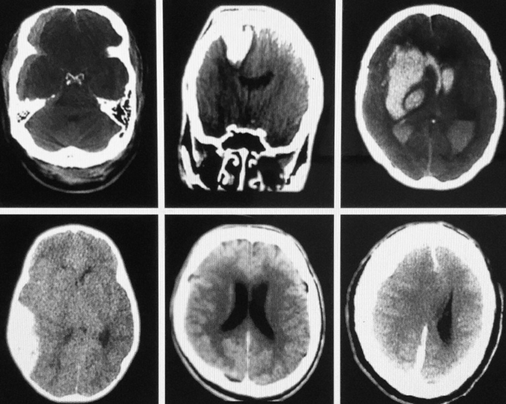
6 CT scans of the brain. Top Lt: Rt SDH w/occipital hematoma
Top middle: Penetrating injury w/round hematoma & surrounding edema.
Top Rt: Hypertensive Intracranial hemorrhage w/extension of blood into the posterior horns of the lateral ventricles bilaterally.
Bottom Lt: Acute Rt. EDH w/shift to Lt. [compression of 3rd ventricle].
Bottom middle: Diffuse global SAH w/convexity block [dilated ventricles].
Bottom Rt: Large acute SDH w/extension along the falx cerebrii, collapse of Rt. ventricle & shift of brain parenchyma to the Lt. From “The Complete EM Prep!” Written/illustrated by Dwight Collman MD. 1192 pages.
